
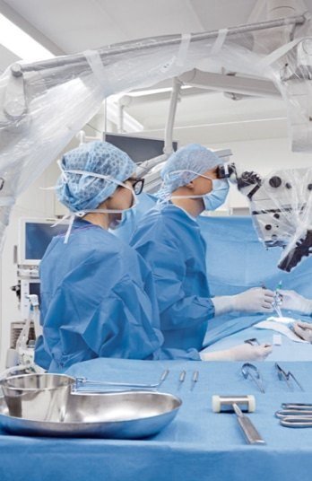
What is a surgical microscope? It is a precision optical instrument, also called an operating microscope, that gives the surgeon a bright, magnified, 3D view of the area they are operating on. It sits on an arm above the patient, so the surgeon has room to work underneath it with instruments.
Neurosurgeons, spine surgeons, ENT surgeons, eye surgeons and plastic surgeons all rely on them. This guide explains how they work in plain language.
Key takeaways
- A surgical microscope magnifies and lights the surgical field while leaving room for instruments.
- Its main parts are the optics head, objective lens, eyepieces, light source, arm and stand.
- Magnification depends on the zoom setting, the eyepieces and the working distance.
- It cannot be sterilized, so it is covered with a sterile drape for every case.
What Is a Surgical Microscope Used For?
Surgeons use a surgical microscope whenever the structures they work on are too small or too deep to see well with the naked eye.
| Specialty | Typical uses |
|---|---|
| Neurosurgery | Brain tumors, aneurysms, nerve decompression |
| Spine surgery | ACDF, microdiscectomy, decompression near the spinal cord |
| ENT | Middle ear surgery, vocal cord work, skull base surgery |
| Ophthalmology | Cataract, retina and corneal surgery |
| Plastic and reconstructive | Joining tiny blood vessels and nerves (microsurgery) |
| Dentistry | Root canals and fine endodontic work |
How a Surgical Microscope Works
Light from the microscope shines down onto the surgical site. That light bounces back up through the objective lens, through a zoom system, and into the eyepieces.
Because there are two light paths (one for each eye), the surgeon sees in 3D. Depth is critical when working near nerves and blood vessels.
The Main Parts of a Surgical Microscope
| Part | What it does |
|---|---|
| Optics head (optics carrier) | Holds the lenses, zoom and focus system |
| Objective lens | The main lens facing the patient. Sets the working distance |
| Eyepieces (oculars) | Where the surgeon looks. Many scopes have a second set for an assistant |
| Light source | LED, xenon or halogen light that brightens the surgical field |
| Arm | Lets the head move and hold its position |
| Stand or mount | Floor stand, ceiling, wall or tabletop |
| Handgrips and foot pedal | Control focus, zoom and movement without touching anything non-sterile |
| Camera (optional) | Records or shows the view on a screen |
How Magnification Works
Total magnification depends on three things:
- The zoom setting. Most surgical microscopes zoom smoothly from a wide view to close detail.
- The eyepieces. Different eyepieces change the final magnification.
- The working distance. This is the distance from the objective lens to the patient. A longer distance gives more room for instruments but changes the field of view.
Surgeons usually start at low magnification to get oriented, then zoom in for fine work.
Quick tip: More magnification is not always better. At high zoom, the field of view gets smaller and depth of focus shrinks. Surgeons balance detail against how much they can see.
Light Sources: Halogen, Xenon and LED
Good light is half of what a surgical microscope does. Three types are common:
| Light source | What to know |
|---|---|
| Halogen | Older technology, warmer light, bulbs need replacing more often |
| Xenon | Very bright, white light, common on neurosurgery scopes, bulbs wear over time |
| LED | Long life, less heat, increasingly common on newer models |
Bulbs and light cables are some of the most common service items. A dim view is often a light issue, not an optics issue.
Fluorescence and Imaging Features
Many modern neurosurgery microscopes can switch into special light modes. With the right dye given to the patient, these modes help the surgeon:
- See blood flow in vessels, for example during aneurysm surgery
- Tell some tumor tissue apart from healthy brain tissue
Built-in cameras can also record the case, stream it to a screen for the team, or send it to a teaching room.
Surgical Microscope vs Loupes vs Exoscope
| Surgical microscope | Surgical loupes | Exoscope | |
|---|---|---|---|
| What it is | Floor, ceiling or wall-mounted optics | Magnifying lenses worn on the head | A camera above the field showing a 3D screen view |
| Magnification | Higher, adjustable zoom | Lower, fixed | Adjustable, digital |
| Light | Strong built-in light | Separate headlight needed | Built-in light |
| Team view | Assistant eyepieces and monitor | Surgeon only | Whole team sees the screen |
| Setup | Needs draping and balancing | Quick | Needs draping and setup |
| Cost | Highest | Lowest | High |
Many surgeons use loupes for opening and closing, then bring in the microscope for the delicate part of the case.
A Short History of the Surgical Microscope
- 1921: Swedish ear surgeon Carl-Olof Nylén used a microscope in the operating room for the first reported time, in Stockholm.
- 1953: Carl Zeiss introduced the OPMI 1, developed with ENT and eye surgeons. It is seen as the forerunner of today’s surgical microscopes.
- 1957: Neurosurgeons began using the operating microscope, starting a revolution in microneurosurgery.
- 1960s: Zoom optics and beam splitters arrived, letting an assistant watch and the case be filmed.
- Today: Microscopes offer motorized controls, fluorescence imaging, 4K cameras and links to navigation systems.
Who Makes Surgical Microscopes?
The best-known makers in US operating rooms include:
- Leica Microsystems: ARveo, M530, M525, M720, M520, PROvido and M320
- Carl Zeiss Meditec: Kinevo, Pentero, Tivato, Vario, Sensera and Movena
- Mitaka: MM51 and MM90
- Haag-Streit: Mainly eye and ENT microscopes
Each brand and model needs its own drape. See our drape compatibility chart to match yours.
Benefits and Limitations
Benefits:
- Clear, magnified 3D view
- Strong, shadow-free light
- An assistant can see the same view
- Recording and teaching
Limitations:
- High purchase and service cost
- Takes floor space and setup time
- Needs a model-specific sterile drape for every case
- Surgeons need training to use it well
How a Surgical Team Uses It
Before the Case
- Check the light, zoom, focus and brakes.
- Balance the arm so the head stays where it is placed.
- Cover the microscope with a sterile drape made for that model.
- Re-balance after draping if the model needs it, and check the full range of motion.
During the Case
- The surgeon moves the microscope with sterile handgrips through the drape.
- Focus and zoom are adjusted with handgrip buttons or a foot pedal.
- An assistant can watch through side eyepieces or on a monitor.
After the Case
- Remove and dispose of the drape.
- Clean the microscope as your facility policy requires.
- Report any issues with light, focus or brakes to your service team.
Why Every Microscope Needs a Sterile Drape
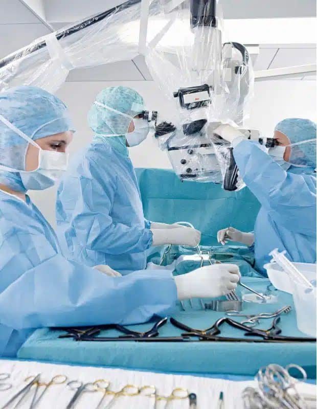
The microscope cannot go through a sterilizer. A sterile drape covers it so the surgeon can touch and move it during surgery without contaminating the field.
The drape must match the microscope model, so the lens window, eyepiece covers and length all fit.
Keeping a Surgical Microscope in Good Shape
Surgical microscopes are expensive and delicate. Regular preventive maintenance keeps the optics, light and brakes reliable.
DB Surgical’s Leica-certified technicians run a 25-point inspection that cleans, lubricates and checks alignment, with a written report after every visit. Learn about microscope maintenance.
Frequently Asked Questions
What is a surgical microscope used for?
It gives surgeons a magnified, well-lit 3D view during delicate surgery, such as brain, spine, ear, eye and microvascular procedures.
How is a surgical microscope different from loupes?
Loupes are magnifying glasses worn on the head. A microscope offers more magnification, better light and a camera option, but takes more space and setup.
Who sets up the surgical microscope?
Usually the circulating nurse and surgical tech prepare and drape it. The surgeon then fine-tunes the position and settings.
Who invented the surgical microscope?
Carl-Olof Nylén first used a microscope in surgery in 1921. Carl Zeiss introduced the first dedicated surgical microscope, the OPMI 1, in 1953.
What light source does a surgical microscope use?
Halogen, xenon or LED. Many newer microscopes use LED for longer life and less heat.
Is an exoscope the same as a surgical microscope?
No. An exoscope uses a camera and a 3D screen instead of eyepieces. It does a similar job in a different way.
How often should a surgical microscope be serviced?
Follow the manufacturer’s schedule. Many facilities book preventive maintenance at least once a year, or more often for busy microscopes.
Related Guides
About American Surgical Products
American Surgical Products has supplied hospitals and surgery centers across the United States since 2010, with no sales reps and no markups. It is the online store of DB Surgical, a Florida medical device distributor founded in 1997 whose Leica-certified technicians service surgical microscopes across the Southeast. Questions? Call 1 (866) 573-7774 or email service@americansurgicalproducts.com. Se habla español.
This article is general information for clinical and purchasing teams. It is not medical advice. Always follow the manufacturer’s instructions for use and your facility’s policies.
Sources
- ASP Preventative Maintenance Visit product page
- DB Surgical: Surgical Microscope Maintenance and Repair
- ASP Surgical Microscope Drape Product Brochure (2024)
- ZEISS: History of microsurgery instruments and the OPMI 1 (1953)
- SPIE: Evolving the surgical microscope (Ma and Fei, Journal of Biomedical Optics 2021)